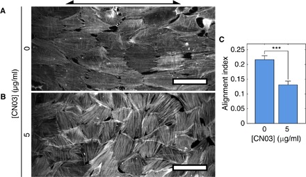
Fig. 6. Rho activation establishes F-actin alignment observed in vivo.

hVSMCs on cylinders with Rc = 125 μm were treated with CN03 [0 μg/ml (A) or 5 μg/ml (B)]. Arrow indicates cylinder axis orientation. Scale bars, 100 μm. (C) AIs of CN03-treated hVSMCs in confluent monolayers on cylinders with Rc = 125 μm. Cells on at least 13 independent cylinders were analyzed in each condition. Results are mean and SE. ***P < 0.001, Student’s t test.
