
Figure 3.
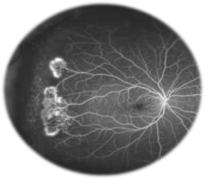
Sea fan formation with neovascularization.
Notes: Fluorescein angiogram image of an individual with sickle cell retinopathy showing sea fan formation with neovascularization. This image was taken using an Optos P200MA ultrawide-field imaging device. This image was originally published in the ASRS Retina Image Bank by Michael P Kelly, FOPS Director, Duke Eye Center Labs, Duke University Hospital. Sickle Cell Retinopathy. 2012; image number, 721. ©American Society of Retina Specialists. http://eyewiki.aao.org/Sickle_Cell_Retinopathy.92