Figure 5.
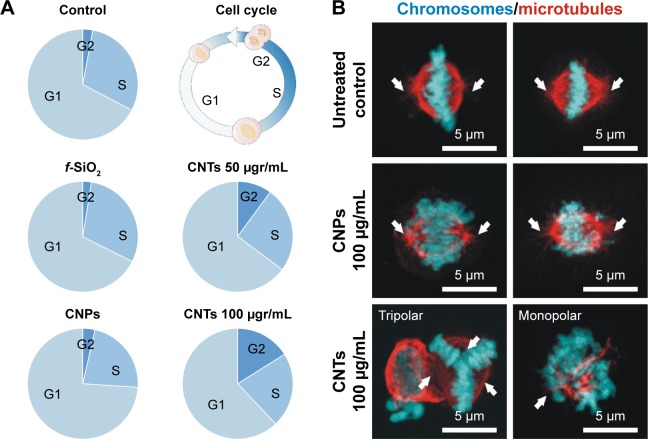
(A) CNPs do not interfere with cell proliferation.
Notes: Quantification of cells at the different stages of the cell proliferation cycle (G1, S, G2) using flow cytometry. CNPs display a similar cell distribution to controls (untreated and f-SiO2-treated cells). Conversely, CNT-treated cells show an obvious dose-dependent cell proliferation blockage in G2. (B) Confocal microscopy projection images of mitotic spindles in untreated controls and cells treated with 100 μg/mL attached to CNPs or dispersed (bottom). Aberrations in the organization of the spindle microtubules (red channel) are observed only in CNT-treated cells. White arrows show the centrosomal location.
Abbreviations: CNPs, CNT-bearing particles; CNT, carbon nanotube; f-SiO2, fluorescent-labeled silica particles.

