
Figure 1. TLR mRNA and protein expression is modulated in response to hyperosmolar stress (HOS) in ocular surface cells.
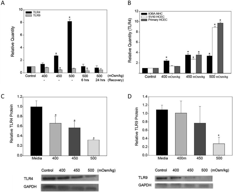
SV40 HCEC were cultured under HOS (400-500 mOsm/kg) or media alone (control) for 24 hrs and following HOS of 500 mOsm/kg, the cells were allowed to recover for 6 and 24 hrs in normal growth media. TLR4 (n=4) and TLR9 (n=3) mRNA expression was determined by quantitative RT-PCR (A). Corneal (primary and SV40 HCEC) and conjunctival epithelial cells (IOBA-NHC) were cultured under HOS for 24 hrs then TLR4 mRNA expression was determined by real-time PCR (B). To confirm a change in protein expression, cell lysates from SV40 HCEC cultured under HOS were analyzed by western blotting for TLR4 (C), TLR9 (D) and GAPDH (n=3). Data were analyzed using an unpaired Student's t-test where P ≤ 0.05 was considered to be statistically significant when compared to control (*).