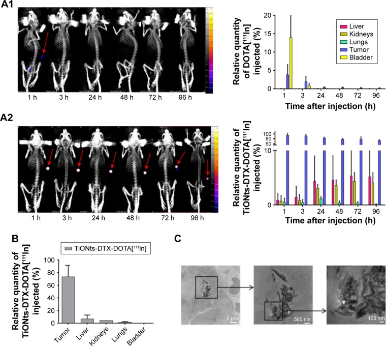
Figure 3.
TiONts-DTX biodistribution analysis.
Notes: (A) SPECT-CT imaging of kinetics and biodistribution analysis for each organ (expressed as a percentage of injected 111In activity, taking into account the decrease in 111In activity after the injection of DOTA[111In] (A1) or TiONts-DTX-DOTA[111In] (A2)). (B) TiONts-DTX-DOTA[111In] biodistribution in dissected organs by radioactivity detection using gamma counting 7 days after injection (mean value ± SD). (C) TEM images showing the intracellular location of TiONts-DTX 24 h after injection into PC-3 tumors.
Abbreviations: DTX, docetaxel; SPECT-CT, single-photon emission computed tomography-computed tomography; TEM, transmission electron microscopy; TiONts, titanate nanotubes.

