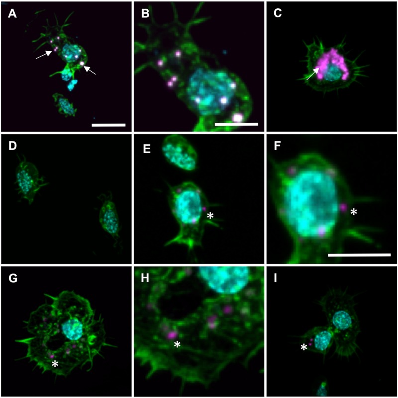
Fig 1. Circulating hemocytes from honey bee’s hemolymph take up the phagocytic markers; latex beads and CM-Dil.
A-C Images showing hemocytes with latex beads (magenta, arrow) detected in the cytosol (labeled with phalloidin in green, nuclei labelled with DAPI in cyan). B is a zoomed image of the same hemocyte (plasmatocyte) detected in A. C Shows a hemocyte (granulocyte) that has internalized multiple latex beads. Such hemocytes were occasionally found. D An example of hemocytes without detectable phagocytosis of latex beads. E-I Hemocytes with detectable CM-Dil (arrow) in the cytosol (green, nuclei labeled with DAPI in cyan). Both F and H are zoomed images of E and G, respectively. E and F Shows a plasmatocyte. G Shows a granulocyte. I The upper cell to the right is an example of a hemocytes without detectable incorporation of CM-Dil, whereas the cell to the left show incorporation of CM-Dil (asterisks). Scale bar for A, C, D, E, G, I in A = 10μm, scale bar for B, F, H in B = 5μm, scale bar in F = 5μm.

