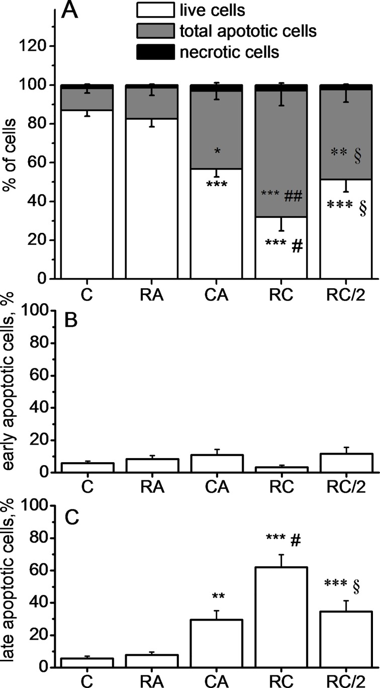
Fig 2.
Quantification of live cells, total apoptotic and necrotic cells (A), early apoptotic cells (B) and late apoptotic cells (C) using annexin V/7AAD staining. Data represent Mean±SD of 8 experiments. Cells were treated with 100 μM RA, 20 μM CA, 100 μM RA + 20 μM CA (RC) or 50 μM RA + 10 μM CA (RC/2). Data are expressed as the percentage of gated cells. ***p<0.001, **p<0.01 and *p <0.05 vs. Control, respectively, ###p<0.001 and #p <0.05 vs CA alone, §p<0.05 vs RC and p>0.05 vs CA alone. Significance signs apply to the corresponding graph bar color

