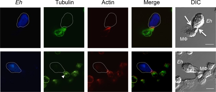
Fig 1. Cytoskeletal modulation in macrophages upon contact with Eh.
Confocal microscopy of THP-1 macrophages incubated with Eh for 10 min. Eh were stained with Cell Tracker Blue prior to incubation. Cells were then fixed, permeabilized and stained with Alexa-488-conjugated anti-tubulin, or isotype control, and with Alexa-633-conjugated phalloidin. Images are representative of three independent experiments. The dotted white line indicates the position of Eh and the arrows in the DIC images shows the point of contact with the macrophage (MΦ). The arrowhead (tubulin frame in lower panel) shows the macrophage microtubule-organizing center. Scale bar 10μm.

