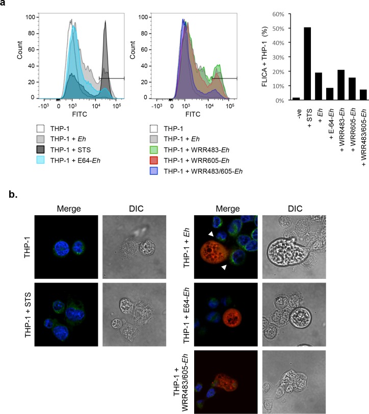
Fig 6. Eh-triggered caspase-6 activation is dependent on EhCP-A1 and EhCP-A4 and is localized at sites of contact.
(a) Caspase-6 activation was measured by flow cytometry with the FITC-labeled caspase-6-FLICA probe in THP-1 macrophages incubated with 10:1 Eh, E64-treated Eh, CP-A5-deficient Eh, WRR483 pre-treated Eh, WRR605 pre-treated Eh, Eh pre-treated with both WRR483 and WRR605, left untreated, or with STS as a positive control. Eh were excluded from analysis based on forward and side scatter. Gating of percent positive cells was established according to the STS-positive population and percent positive cells for each treatment are indicated in the accompanying graph. (b) Caspase-6 activation for each treatment was also visualized by confocal microscopy using caspase-6-FLICA (green), DAPI (blue). Eh were pre-treated with Cell Tracker Orange (red) prior to incubation with THP-1 macrophages. STS was used as a positive control. Arrowheads show areas of polarization of caspase-6 towards the Eh-macrophage interface. The dotted white line indicates the position of Eh and the point of contact with the macrophage (MΦ). Results are representative of three independent experiments.

