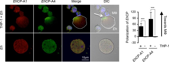
Fig 7. Cell surface EhCP-A1 and EhCP-A4 polarize to the macrophage contact site.
To assess EhCP-A1 and EhCP-A4 localization, Eh were incubated with DAPI-treated THP-1 cells (top panels) or not (bottom panel). Slides were then stained with EhCP-A1 and EhCP-A4, and the appropriate secondary antibodies. EhCP-A1 is shown in red, EhCP-A4 is shown in green. Both EhCP1 and EhCP4 polarized on the Eh surface towards the macrophage whereas no polarization was observed in Eh basally (bottom panel). The dotted white line indicates the position of Eh and the arrow in the DIC images shows the point of contact with the macrophage (MΦ). Polarization of EhCPs was quantified by comparing density of EhCP staining at the leading half facing the macrophage in comparison to the tailing half. ***p<0.001 as determined by Student’s t-test.

