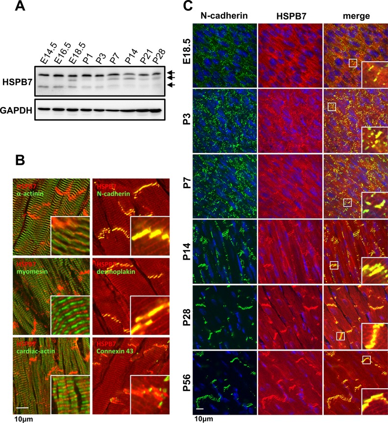
Fig 1. Expression and localization of HSPB7 in cardiac muscle.
(A) Immunoblot analysis of the cardiac muscle showing HSPB7 constitutive expression from the embryonic stages to adulthood (E14.5 to P28) with multiple forms of HSPB7 at different molecular masses (arrows). (B) Subcellular localization of HSPB7 in the cardiac muscle of adult mice. The heart sections were stained with antibodies against HSPB7 (red) and desmoplakin (desmosome), α-actinin (Z-line), myomesin (M-line), N-cadherin (adhering junction), connexin 43 (gap junction), and cardiac-actin (I-bend). HSPB7 mainly localizes at the intercalated discs and is adjacent to the Z-line with a striated pattern. (C) Colocalization of HSPB7 with N-cadherin during development. Heart sections from the embryonic stages to adulthood (E14.5 to P56) were stained with antibodies against HSPB7 (red) and N-cadherin (green). The nucleus was visualized through Hoechst 33342 staining. Insets show the representative areas with higher magnification. Scale bar: 10 μm.

