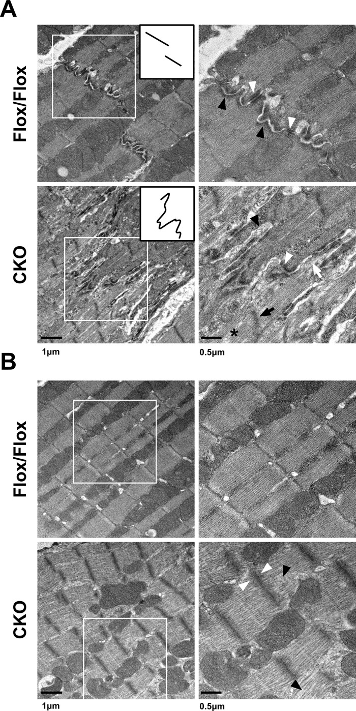
Fig 5. Ultrastructural study of control and HSPB7 CKO hearts.
Transmission electron micrographs (TEMs) of ventricular myocardium from HSPB7 CKO and control mice at d7 after tamoxifen administration. Right panels are higher-magnification views of the boxed areas in the left panels. (A) Normal intercalated disc structures were visible in the control hearts. The inset provides a simplistic representation of the morphology of the intercalated discs. Higher-magnification images showed abnormal adherens junctions (black arrowheads) and desmosomes (white arrowheads) with widened gaps of the intercalated discs in HSPB7 mutant hearts. Abnormal Z-line (black arrow), filament disruption (asterisk), and detachment of myofibrils at the intercalated disc (white arrow) were also observed in the CKO mice. (B) The ultrastructure of the sarcomeres at the center part of the cardiomyocyte was slightly distorted. Higher-magnification images showed (right panel) loose actin filaments (black arrowhead) and wider, less dense Z-lines (white arrowhead) compared with the controls. n = 3 per group. Scale bar: 250 nm.

