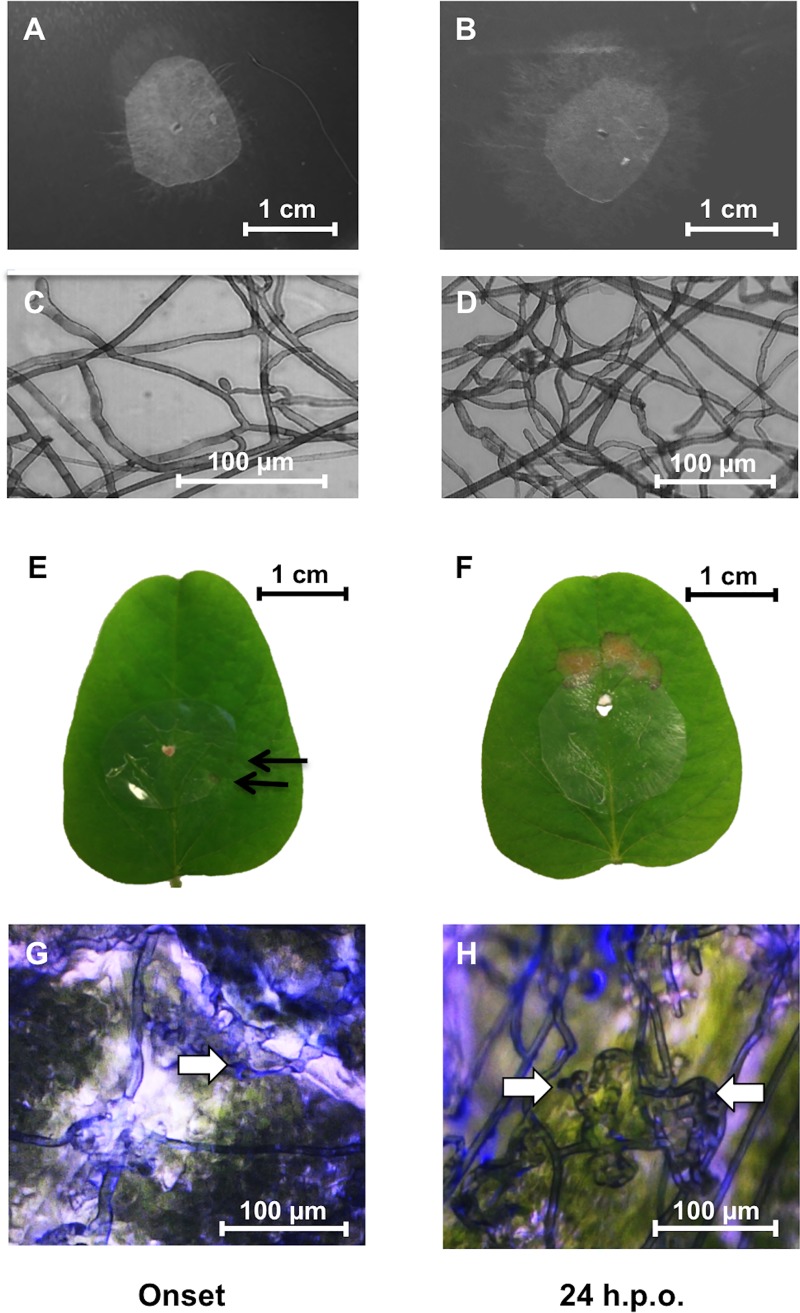
Fig 2. Rhizoctonia solani-soybean interactions at onset and 24 h.p.o. of necrosis.
In vitro controls on PDA harvested at onset (A) and 24 h.p.o. (B) of necrosis. Microscope images of hyphae on nitrocellulose membranes in vitro at onset (C) and 24 h.p.o. (D) of necrosis showing normal growth and lack of hyphal aggregates and infection structures. Soybean leaf samples infected with R. solani AG1-IA at onset (E) and 24 h.p.o. (F) of necrosis. Arrows indicate the onset of necrotic lesions approximately 36 h post-inoculation. Microscope images of hyphae on nitrocellulose membranes from soybean leaves at onset (G) and 24 h.p.o. (H) of necrosis showing infection cushion structures (arrows). Note that hyphal aggregation and infection cushion initials occurred only in R. solani samples grown on membranes overlaid on leaves (G, H) and not on PDA (C, D).

