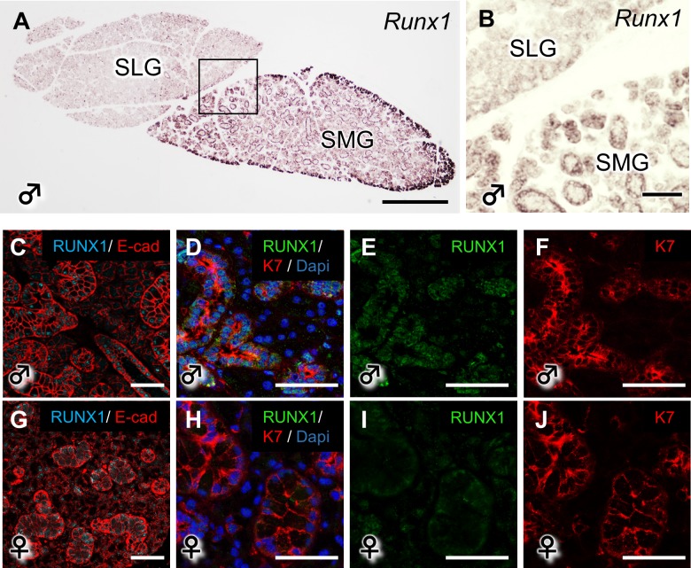
Fig 1. The expression of RUNX1 in the mouse salivary glands.
(A, B) The mRNA expression of Runx1 in the SMG and SLG of P50 WT mice. B shows a higher magnification view of A. Scale bars: 500 μm (A), 100 μm (B). (C-J) The RUNX1 protein expression on P50 in the WT SMG is shown by anti-RUNX1 staining under a confocal microscope. Scale bars: 100 μm.

