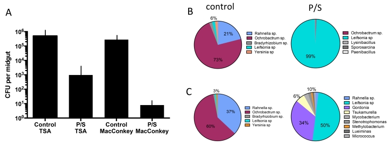
Figure 2.
Changes in the size and diversity of bacterial communities in P. duboscqi following infection and treatment with P/S. Flies were artificially fed through a membrane on mouse blood seeded with 4×10⁶/ ml LmRy promastigotes and treated or not with P/S in the blood and sugar meals. (A) Dissected midguts at 14 days p.i. were cultured on TSA and MacConkey agar. Bar graphs show mean CFUs + SD, 10 midguts/group. (B) Culture dependent, and (C) culture independent analysis of the gut bacteria at 14 days p.i. based on PCR amplification and cloning of the 16S DNA prepared from pools of 10 flies in each group.

