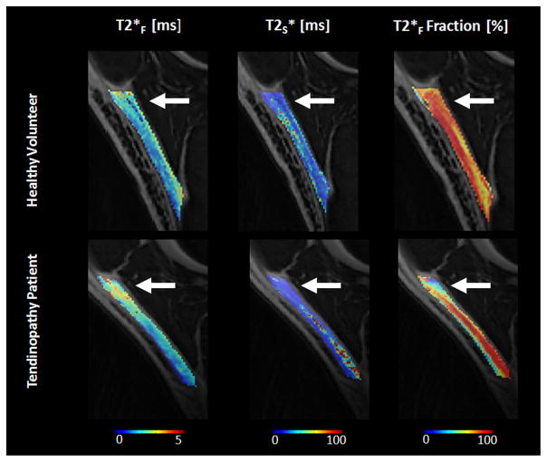
Figure 2.
UTE-T2* parameter maps in a 25 year old healthy male volunteer and a 21 year old high-level collegiate male basketball player with grade 0 patellar tendinopathy (i.e. clinically diagnosed disease without associated MRI findings of tendon thickening and increased signal intensity). Note the small focal area of increased T2F and decreased FF in the proximal patellar tendon in the patient with patellar tendinopathy with no visible change in T2S (arrows).

