
Fig 4. SC (negative control) ISH.
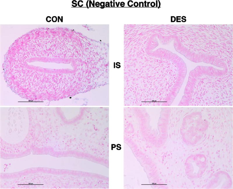
Shown here and in the following micrographs (Figs. 5 – 12) are representative results for cross sections of control (CON) or neonatally DES-exposed uteri from IS and PS animals following ISH processing with the indicated RNA probe and then staining with fast red nuclear counterstain. After incubation with the scrambled negative control probe (SC), note here the uniformly pink-counterstained nuclei and lack of blue reaction product in all four tissue sections.