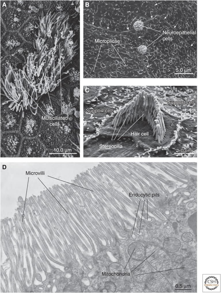
Figure 1.
Survey of apical surface protrusions found in epithelial cells. (A) Scanning electron micrograph (SEM) of the apical surface of the rat trachea showing multiciliated cells. Adjacent cells are nonciliated or have rudimentary cilia. (B) SEM of the mucosal surface of the rat proximal urethra. Microplicae are observed at the apical surface of umbrella cells found in this region. Arrows mark the junctional ring of adjacent cells. The apical surfaces of neuroepithelial cells, which are covered by an apical tuft of short microvilli, are interspersed between adjacent cells. (C) SEM of the cochlea of the adult mouse showing a hair cell with associated “hair bundle,” which is comprised of stereocilia arranged in a stair-step configuration. The apical surfaces of adjacent support cells are studded with microvilli. (D) Transmission electron micrograph of microvilli at the apical surfaces of the rat proximal tubule epithelial cells. Endocytic pits and mitochondria are marked. (Electron micrograph in panel C was kindly provided by Jonathan Franks, Center for Biological Imaging, University of Pittsburgh; and micrographs in panels A, B, and D were kindly provided by Wily G. Ruiz, Kidney Imaging Core, University of Pittsburgh.) (Figure is from Apodaca and Gallo 2013; adapted, with permission, from the authors.)

