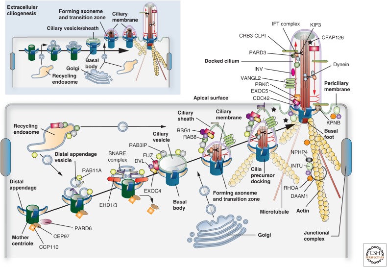
Figure 7.
Models for ciliogenesis. Cilia are formed by one of two mechanisms: “intracellular ciliogenesis” or “extracellular ciliogenesis.” The former is depicted in the larger panel and the latter in the smaller panel found in the upper left of the figure. Both should be viewed from left to right. The proteins and associated binding partners depicted in this figure are culled from numerous studies, and may vary depending on organism, cell type, and mechanism of ciliogenesis. (Larger panel) In “intracellular ciliogenesis,” a cilia precursor is formed in the cytoplasm and is subsequently delivered to the apical surface where it undergoes docking. Initially, a mother centriole with attached distal appendages accumulates RAB11A-positive distal appendage vesicles, which subsequently recruit EHD1/3, triggering a fusion event that results in the formation of a “ciliary vesicle.” Concomitantly, the centriole discharges CEP90 and CCP110, marking the conversion of the centriole to the basal body. Either before, or subsequent to ciliary vesicle formation, FUZ recruits DVL, the exocyst subunit EXOC4, and eventually RSG1 to the vesicle surface. In a similar fashion, RAB11 recruits the RAB8 exchange factor RAB3IP. In the next step, protein components from the Golgi (e.g., IFT20, ciliary membrane proteins, and PCP core proteins such as VANGL2), from recycling endosomes (e.g., RAB8), and from the cytoplasm (e.g., IFT and transition zone proteins) arrive at the ciliary vesicle and begin to assemble the axoneme and transition zone. This step is marked by the presence of the ciliary sheath, which will ultimately give rise to the periciliary membrane, and covers the inner ciliary membrane. Expansion of both membranes likely requires continued input from endosomes and Golgi. The ciliary precursor undergoes fusion in a process called “docking,” followed by further building of the axoneme and transition zone. The cilium is associated with both microtubules and actin filaments. (Smaller panel) Extracellular ciliogenesis is observed in cells with multiple cilia. Here, the centriole is delivered to a position just below the apical surface, undergoes maturation, and a ciliary vesicle/sheath is formed. The latter undergoes fusion with the apical plasma membrane, a step that is followed by assembly of the transition zone and axoneme at the cell surface.

