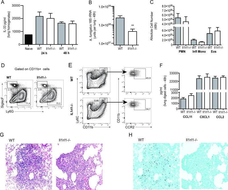
Figure 1. The lack of IL-33 signaling results in enhanced A. fumigatus lung clearance.
(A) C57BL/6 wild-type and Dectin-1 deficient (Clec7a−/−) mice were challenged intratracheally with A. fumigatus conidia (Af) and 48 h post-exposure, whole lungs were collected and IL-33 levels quantified in clarified lung homogenates by ELISA. The Figure illustrates cumulative data from two to three independent studies (n = 3–5 mice per group, per study). (B) C57BL/6 wild-type (WT) and Il1rl1−/− (IL-1RL1; ST2 deficient) mice were challenged intratracheally with A. fumigatus conidia and 48 h after exposure, lung fungal burden was assessed by real-time PCR analysis of A. fumigatus 18S rRNA levels. The Figure illustrates cumulative data from two independent studies (n = 4–5 mice per group, per study). Data are expressed as mean A. fumigatus 18S rRNA + SEM. (C) Cumulative flow cytometric data from two independent studies (n = 2–4 mice per group, per study). Data are expressed as absolute number of live cells in lung digests. (D/E) Lung cells were isolated via enzymatic digestion, Fc-blocked, stained with a live/dead staining kit and thereafter stained with fluorochrome-conjugated CD11c, CD11b, Ly6G, Ly6C and Siglec F. Representative flow cytometric plots are included for (D) neutrophils and eosinophils (gated on CD11b+ cells followed by Ly6G+ cells as neutrophils and Siglec-F+ cells as eosinophils) and (E) inflammatory monocytes (gated on CD11b+ Ly6C+ cells followed by gating on CCR2+ cells). (F) C57BL/6 wild-type (WT) and Il1rl1−/− (IL-1RL1; ST2 deficient) mice were challenged intratracheally with A. fumigatus conidia and 48 h after exposure, the right lungs were collected, enzymatically digested and unfractionated lung cells cultured in triplicate for 24 h. CCL11/eotaxin, CXCL1/KC and CCL3/MIP-1α levels were quantified in clarified co-culture supernatants by Bio-Plex. The Figure illustrates cumulative data from four independent studies (n = 1–2 mice per group, per study). (G) Representative H&E-stained lung sections from wild-type (left) and Il1rl1−/− (right) mice challenged intratracheally with A. fumigatus conidia for 48 h. Original magnification of 200X. (H) Representative GMS-stained lung sections from wild-type (left) and Il1rl1−/− (right) mice challenged intratracheally with A. fumigatus conidia for 48 h. Original magnification of 200X. For all graphs, ** represents a P value of < 0.01, respectively (Unpaired two-tailed Student’s t test).

