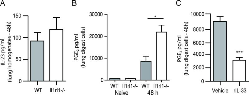
Figure 3. PGE2, but not IL-23, is increased in the absence of IL-33 signaling.
(A) C57BL/6 wild-type (WT) and Il1rl1−/− (IL-1RL1; ST2 deficient) mice were challenged intratracheally with A. fumigatus conidia and 48 h after exposure, whole lungs were collected and IL-23 levels quantified in clarified lung homogenates by ELISA. The Figure illustrates cumulative data from two independent studies (n = 4–5 mice per group, per study). (B) C57BL/6 wild-type (WT) and Il1rl1−/− (IL-1RL1; ST2 deficient) mice were challenged intratracheally with A. fumigatus conidia and 48 h after exposure, the right lungs were collected, enzymatically digested and unfractionated lung cells cultured in triplicate for 24 h. PGE2 levels were quantified in clarified co-culture supernatants by EIA. The Figures illustrates cumulative data from three independent studies (n = 1–2 mice per group, per study). (C) C57BL/6 wild-type mice were challenged with A. fumigatus and 6 and 24 h thereafter, administered IL-33 (1 µg in 50 µl) or PBS intratracheally. Forty-eight h after exposure, the right lungs were collected, enzymatically digested and unfractionated lung cells cultured in triplicate for 24 h. PGE2 levels were quantified in clarified co-culture supernatants by EIA. The Figure illustrates cumulative data from three independent studies (n = 1–2 mice per group, per study). For all graphs, * or *** represent a P value of < 0.05 or 0.001, respectively (Unpaired two-tailed Student’s t test).

