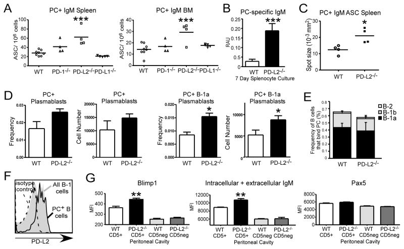
Figure 3. PD-L2 deficiency results in increased PC-specific ASC numbers in the spleen and bone marrow.
A) ELISPOT analysis of PC-specific IgM+ ASC number in the BM and spleen of naïve WT, PD-1−/−, PD-L2−/−, and PD-L1−/− mice. B) ELISA analysis of PC-specific IgM in supernatant from unstimulated, naïve 7-day splenocyte cultures. C) ELISPOT analysis of splenic PC-specific IgM+ ASC size in WT and PD-L2−/− mice. D) Frequency and number of splenic PC-specific total plasmablasts and PC-specific B-1a plasmablasts (CD5+19+138+) in WT and PD-L2−/− mice. E) Frequencies of peritoneal B cells (CD19+CD11b+/−) that bind PC in WT and PD-L2−/− mice, with distribution among B-1a, B-1b, and B-2 cells indicated. F) PD-L2 expression on PC-specific peritoneal B cells in WT mice relative to total B-1 cell PD-L2 expression and isotype control staining. G) Blimp1, total IgM (intracellular + extracellular), and Pax5 levels in peritoneal B cells (CD5+19+ vs CD5neg19+) in WT and PD-L2−/− mice. N ≥ 4 mice/group; for A, analyzed by ANOVA with Dunnett Post Test; B-G analyzed by Student’s t test (* p<0.05, ** p<0.005, *** p<0.001).

