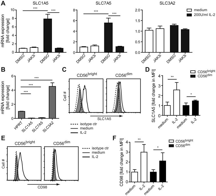
Figure 2. IL-2 priming increases the expression of SLC1A5 and CD98 in NK cells.
(A,B) NK cells were enriched from PBMCs and treated with DMSO (0.05%) or 500 nM Jak3i for 2 h. Cells were then cultured in medium ± 200 U/ml IL-2 for an additional 6 h. RNA was isolated and mRNA expression was analyzed by real-time PCR. (A) Show fold change of mRNA expression relative to the DMSO sample of the medium treated cells, calculated by ΔΔCT method, from three donors. (B) Show fold change of mRNA expression relative to the HPRT sample from three donors. (C-F) Enriched NK cells were cultured in medium ± 200 U/ml IL-2 for 24 h. (C, D) The intracellular SLC1A5 protein level and (E, F) surface expression of CD98 on CD56bright and CD56dim NK cells was measured by flow cytometry. (C, E) Results are shown for one donor and are representative of four donors. (D, F) Data show fold change in mean fluorescence intensity (MFI) ± s.d. relative to the average MFI of the medium treated samples from four donors. Statistical analysis was performed by 2-tailed unpaired student's t-test (*p<0.05, **p<0.01, ***p<0.001).

