
Figure 5. IL-6R antibodies inhibit STAT3 activation, decrease tumor cell proliferation, and sensitize tumors to chemotherapy-induced apoptosis.
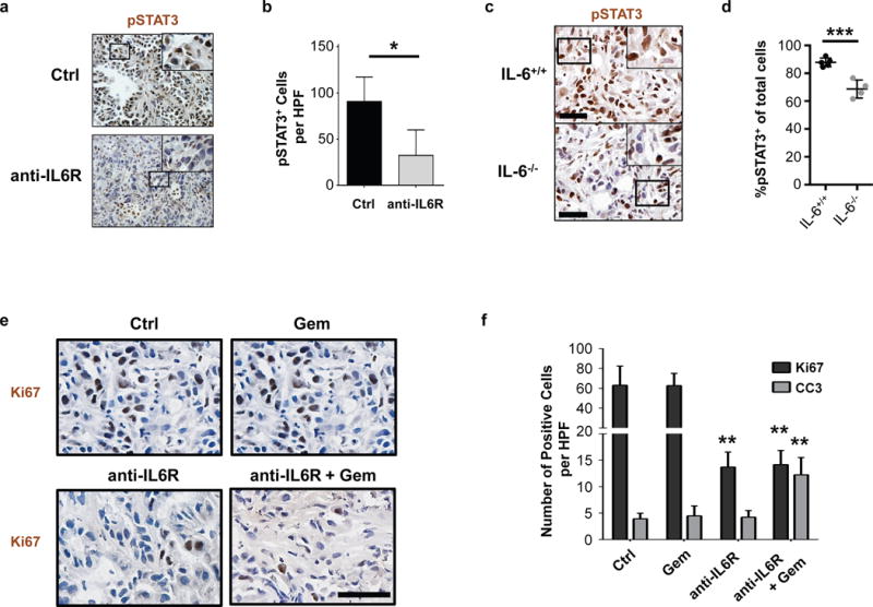
(A) Representative images showing immunohistochemical staining for pSTAT3 expression in tumor tissue from KPC mice treated with anti-IL6Ralpha blocking antibodies or isotype control. Scale bar = 100μm. (B) Quantification of pSTAT3+ cells detected in (A) by immunohistochemistry. n=3–4 mice per group, 4 high-powered fields per mouse; *, p<0.05; two-tailed student’s t-test. (C) Representative images showing immunohistochemical staining for pSTAT3 expression in tumor tissue from PDAC cell line implanted orthotopically into syngeneic wild-type (IL-6+/+) or IL-6 deficient (IL-6−/−) mice. (D) Quantification of the percentage of pSTAT3+ cells per total nucleated cells detected in (C) by immunohistochemistry. n=4–5 mice per group, 5 high-powered fields per mouse. ***, p<0.001; two-tailed student’s t-test. (E) Tumor-bearing mice were treated with or without anti-IL-6Ralpha for three days. Shown are representative images of Ki67 expression in PDAC tumors isolated one day after subsequent treatment with or without gemcitabine chemotherapy. Scale bar = 50μm. (F) Quantification of Ki67 and cleaved caspase 3 (CC3) detected in PDAC tumors isolated one day after gemcitabine chemotherapy treatment with or without three-day pretreatment with anti-IL-6Rα antibodies. n=4–5 mice per group, 5–8 high-powered fields per mouse. Statistical significance determined using two-tailed student’s t-test. Comparisons were made to Ctrl. **, p<0.01.