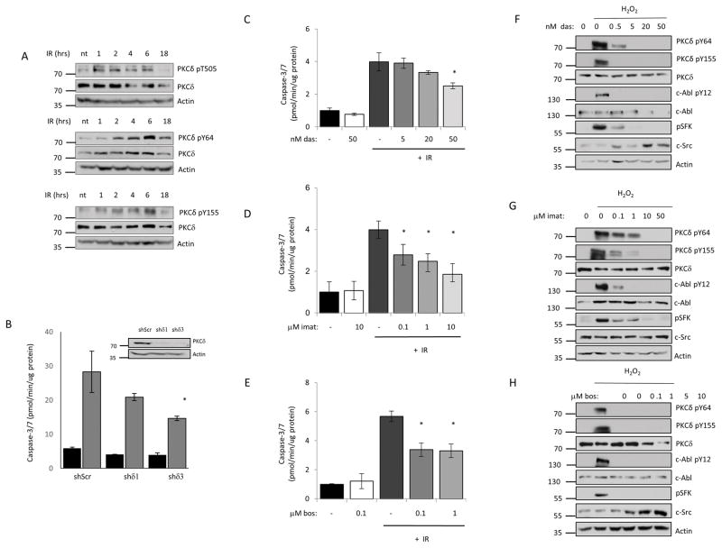
Figure 1. Tyrosine kinase inhibitors suppress tyrosine phosphorylation of PKCδ and IR induced apoptosis in salivary gland acinar cells.
A, ParC5 cells were treated with 10 Gy IR and collected at the indicated time after IR. Whole cell lysates were resolved with SDS-PAGE and analyzed using phospho-specific antibodies that recognize PKCδ pY64, pY155 or pT505. Membranes were stripped and probed for total PKCδ and actin to determine loading. Each experiment was done a minimum of three times; representative immunoblots are shown. B, ParC5 cells were transiently infected with lentivirus expressing PKCδ specific shRNAs (shδ1 or shδ3) or a scrambled control shRNA (shScr), after 72 hours treated with IR (10Gy), and harvested after an additional 18 hr. Caspase-3/7 activity was assayed as described under “Methods”. Data shown is the average of triplicate samples from a representative experiment plus and minus the S.D. (error bars). The experiment was repeated three times, * = p<0.05 for caspase activity compared to cells expressing shScr. An immunoblot showing depletion of PKCδ is shown below. For panels C–H: ParC5 cells were treated with increasing concentrations of dasatinib (C, F), imatinib (D, G), or bosutinib (E, H) for 30 min, followed by treatment with 10 Gy IR (C–E), or the addition of 5 mM H2O2 for 30 min (F–H). C–E, Lysates were collected for caspase-3/7 activity analysis 18 hours after IR and was assayed as described under “Methods.” The data are the average of triplicate measurements from a representative experiment plus the S.D. (error bars). Each experiment was repeated three times. * = p<0.05 for caspase activity compared to cells treated with IR alone. F–H, Whole cell lysates were collected 30 minutes after H2O2 treatment and analyzed by immunoblot for PKCδ pY64 and pY155. Inhibition of c-Src and c-Abl was determined by probing for their respective activation sites pY416 (c-Src) and pY412 (c-Abl). Blots were stripped and probed for total PKCδ, total SFK, total c-Abl and actin to determine total protein levels and loading efficiency. Each experiment was done a minimum of three times; representative immunoblots are shown.

