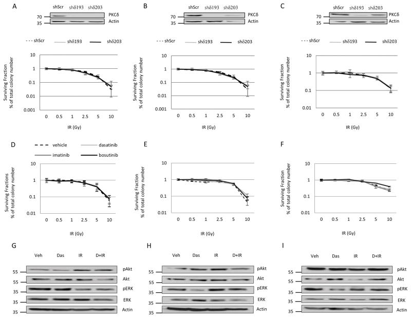
Figure 4. Depletion of PKCδ or treatment with TKIs does not enhance survival of HNSCC cells.
A–C, Cal27 (A), FaDu (B), or UMSCC19 (C) HNSCC cells were stably transduced with lentivirus to express PKCδ specific shRNAs (shδ193 and shδ203) or a scrambled control (shScr). An immunoblot showing depletion of PKCδ is shown above. 24 hours after plating, cells were exposed to IR (0–10Gy). D–F, Cal27 (D), FaDu (E), and UMSCC19 (F) cells were plated, and 24 hours later were treated with DMSO (vehicle), dasatinib (50 nM), imatinib (10 μM), or bosutinib (1 μM) for 30 min prior to IR (0–10 Gy). A–F, Colonies were allowed to form over 10–14 days. The plating efficiency and surviving fractions from each condition was quantified using Image J as described in Methods. The data represents the average plating efficiency from three independent experiments plus the standard deviation (error bars). G–I, Cal27 (G), FaDu (H), and UMSCC19 (I) cells were treated with DMSO (Veh), 50 nM dasatinib alone (Das), or 10 Gy IR alone (IR), or given 50 nM dasatinib 30 min prior to 10 Gy IR (D + IR); cells were harvested 1 hour after IR. Whole cell lysates were analyzed for phospho-Akt and phospho-ERK, and stripped and probed for total Akt, total ERK, and actin. Shown are representative images from one experiment that was repeated three or more times.

