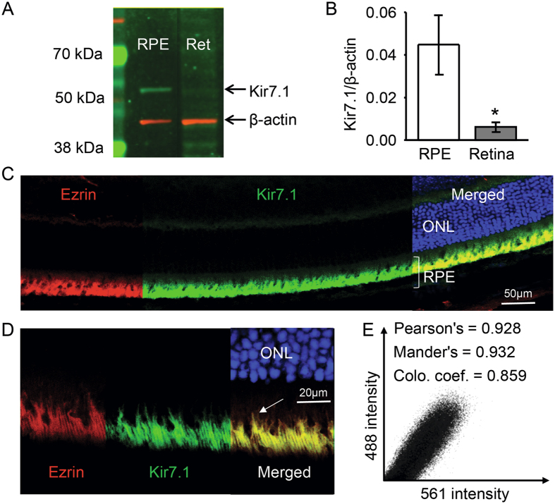
Figure 2.
Kir7.1 protein expression. (A) Protein expression of Kir7.1 in RPE and retinal tissue detected by Western blot analysis. Complete Western blot image is included as Supplemental Fig. 2A. (B) Bar graph showing the relative expression of Kir7.1 protein in the RPE and the retina by densitometry expressed as a Kir7.1/b-actin ratio. (C) Immunohistochemistry against Kir7.1 and Ezrin; ezrin (red) labels the microvilli of the RPE cells and Kir7.1 (green) co-localizes with ezrin confirming its presence in the apical processes of the RPE cell. Outer nuclear layer (ONL) is stained with DAPI (blue). Scale bar is indicated. A video is included as a Supplemental data. (D) Higher magnification image of the RPE layer shows ezrin expression (red) in apical processes extending towards the retina. Kir7.1 (green) staining also appeared in apical membrane extensions with co-localization of both proteins (yellow) in long apical processes (arrow). Scale bar is included. (E) A quantitative distribution plot representation of signals acquired in green (488) vs. red (594).

