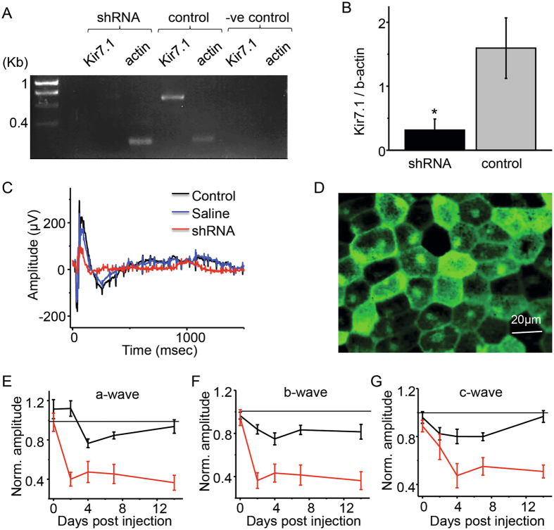
Figure 3.
Reduced RPE/retina function after sub-retinal administration of lentivirus containing shRNA for Kir7.1. (A) Gel electrophoresis showing that Kir7.1 mRNA expression is reduced after Kir7.1 shRNA injection when compared to the un-injected contralateral eye. Full image of the gel is included as Supplemental Fig. 3A. (B) Expression of Kir7.1/b-actin ratio in shRNA injected and un-injected eye (part A) represented in a bar graph. (C) Representative scotopic traces comparing the shRNA-injected mice 14 d post injection, as well as control and PBS-injected mice. (D) Fluorescent image of live RPE sheet of cells displaying the expression of GFP that is fused with the shRNA Kir7.1. (E) Normalized a-wave and (F) normalized b-wave comparing the shRNA injected eye with the PBS injected eye at 30 cd.s/m2 at 0, 2, 4, 7, and 14 d post-injection. (G) Comparison of c-wave for eyes injected with Kir7.1 shRNA and PBS at 0, 2, 4, 7 and 14 d post injection. Dotted line represents the control-normalized response from non-injected eyes.

