Figure 6.
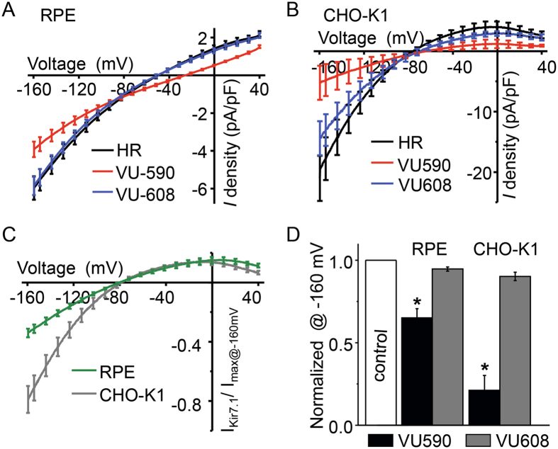
Inhibition of Kir7.1 current by VU590. Representative traces of whole cell currents recorded from single isolated RPE cells from mouse (A) and CHO-K1 cells (B) overexpressing the Kir7.1 channel, respectively. Whole cell currents were recorded by voltage ramping from +40 mV to −160 mV. The traces represent current density, which is the whole cell current normalized for the cell capacitance. In both cell types, the normal baseline Kir7.1 current is recorded prior to cells being treated with 50 µM VU590. The current is reduced after treatment with VU590, but is not affected by the treatment with 50 µM VU608. (C) and (D) The normalized Kir7.1 current at −160 mV is compared before, and after, treatment with VU590 or VU608.

