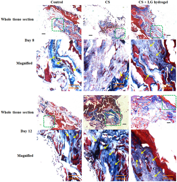
Figure 6.
Masson’s trichrome staining. Synthesis and deposition of collagen were evaluated using Masson’s trichrome stained granulation tissues of days 8 and 12. The boxed regions in the whole tissue sections (4x; scale bar 25 µm) are displayed at higher magnification (20x; Scale bar 100 µm) as magnified images. CS+LG hydrogel treated tissues showed enhanced collagen synthesis, deposition, and maturation on days 8 and 12, compared to control and CS hydrogel treatment. Yellow arrows indicate the deposition of collagen bundles. C denotes the collagen fibers in treated tissues.

