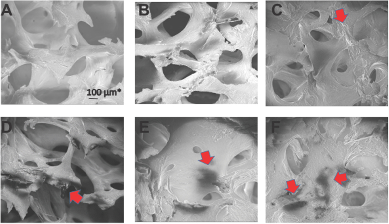
Figure 6.
Representative Scanning Electron Microscopic (SEM) images of control and AVNFH bone. (A–C) SEM images of control bone showing honey comb shaped trabecular arrangement. Regions of bone remodelling is also observed (red arrow). (D–F) SEM images of AVNFH bone showing perturbed trabecular arrangement. Trabeculae lose connectivity altering the honey comb shape. In addition, regions of dead bones are observed. (Black areas indicated by red arrow). Extensive remodelling of the bone is indicated by red arrows.

