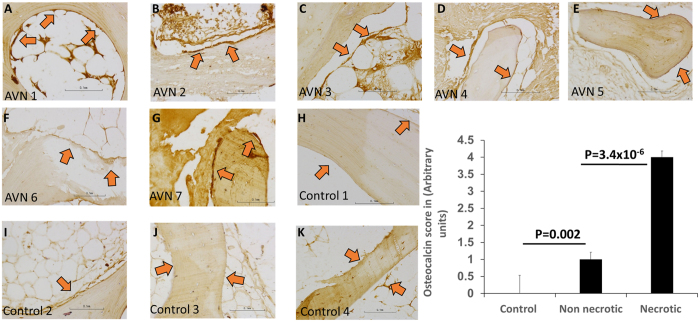
Figure 7.
Photomicrographs showing osteocalcin staining in trabecular bone sections from AVNFH patients (panels A–G) and control individuals (panels H-K). Osteocalcin staining in osteoblasts lining (red arrows) the bone were quantified (panel L) by an expert bone pathologist. Overall, strong osteocalcin staining was observed in osteoblasts lining the necrotic areas in AVNFH sample, indicating extensive bone remodelling (panel L). In contrast, osteoblasts lining the control bone showed weak osteocalcin staining (panel L). Interestingly, within the AVNFH osteocalcin (panel L, refer Supplementary Figure 22). In control bones (derived from sites of fracture), osteoblasts proximal to late stage fracture callus showed strong osteocalcin staining, also suggesting extensive bone remodelling (refer Supplementary Figure 22). In control bones (derived from sites of fracture), osteoblasts proximal to late stage fracture callus showed strong osteocalcin staining, also suggesting extensive bone remodelling (refer Supplementary Figure 22). In all cases, non-specific staining was observed in the marrow. All images photographed at 200X (refer to scale in the Inset).

