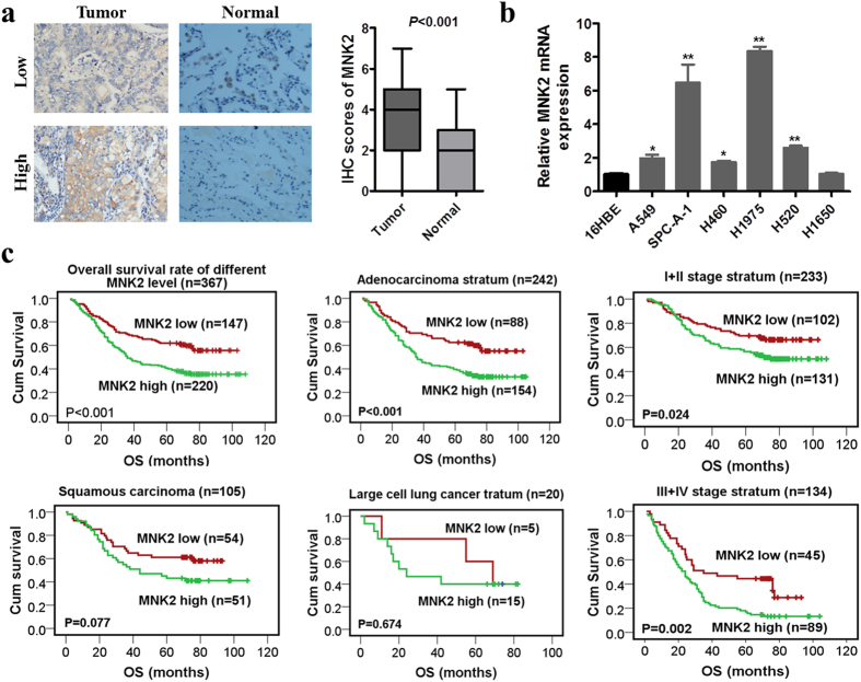
Figure 1.
MNK2 was overexpressed in NSCLC and correlated with poorer prognosis. (a) MNK2 was highly expressed in NSCLC tissues compared with normal adjacent lung tissues (NATs) (**P < 0.01). Representative IHC staining of MNK2 in 2 pairs (low expression and high expression) of NSCLC cases (original magnification: ×200). (b) RT-PCR analysis showed that MNK2 was overexpressed in most NSCLC cell lines compared with normal bronchi epithelial cell line (16HBE) (*P < 0.05, **P < 0.01). (c) Kaplan-Meier analysis showed that the overall survival (OS) rates of NSCLC patients with MNK2 overexpression were lower, especially in adenocarcinoma and clinical stage III + IV (P < 0.01).

