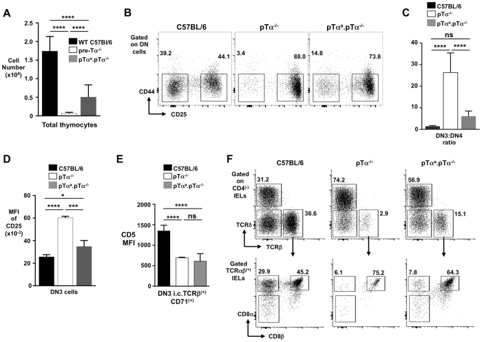
Figure 3.
Reduced unconventional TCRαβ(+)CD8αα(+) IELs in pTαa-only transgenic mice. (A) Summary bar chart of DP cell numbers in C57BL/6, pTα-deficient and pTαa. pTα−/−mice. (B) Representative flow cytometry profiles of DN3 and DN4 subsets from thymus of C57BL/6, pTα−/− and pTαa. pTα−/− transgenic mice. (C) Bar chart summarizing the DN3 to DN4 ratio in C57BL/6, pTα-deficient and pTαa. pTα−/−mice. (D) Summary bar chart of MFI of CD25 on DN3 thymocytes from each strain of mice. (E) Bar chart showing CD5 MFI on β-selected DN3 cells (i.c.TCRβ(+)CD71(+)) from each strain of mice. (F) Representative flow cytometry plots (from n > 6) of IEL populations (TCRαβ(+)CD8αα(+) and TCRαβ(+)CD8αβ(+) IELs) from the small intestine of C57BL/6, pTα-deficient and pTαa. pTα−/− mice. Percentages of cells are indicated near each gate. ****p < 0.0001, ***p < 0.001, *p < 0.05, ns is for not significant.

