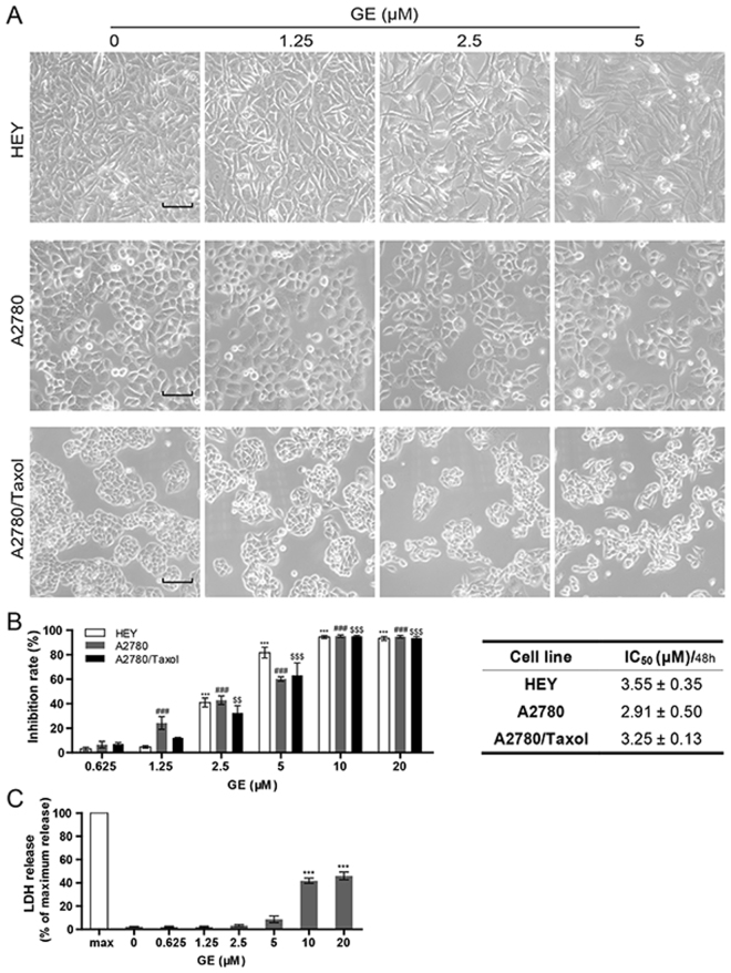
Figure 2.
GE inhibited cell proliferation in ovarian cancer cells. (A) Morphological changes of HEY, A2780, and A2780/Taxol after 24 h of GE treatment. The length of the scale bar is 50 μm. (B) After 48 h of GE treatment, the inhibition rates of cell proliferation of three cell lines were tested by MTT assay. (C) The relative extracellular LDH concentration of HEY cells was tested by LDH release assay. All experiments were performed three times.

