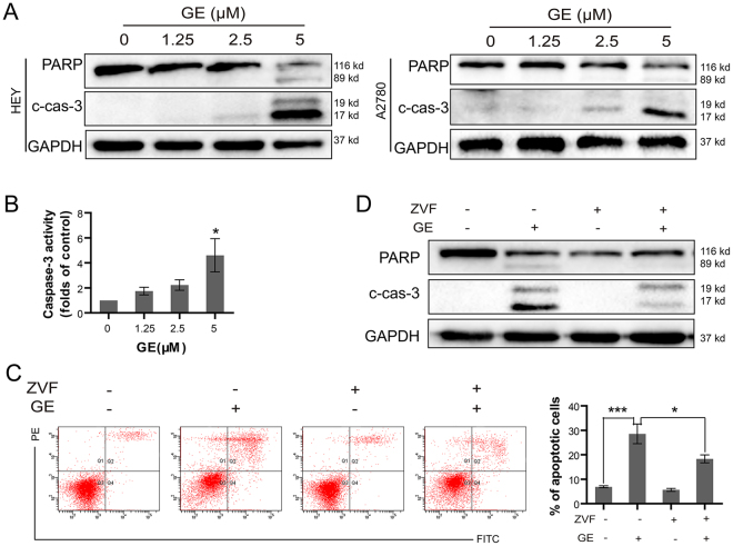
Figure 4.
GE induced apoptosis via caspase signaling pathway. After 24 h of GE treatment, the apoptosis-associated protein in HEY and A2780 cells were examined by western blot (A), and caspase-3 activity in HEY cells was tested (B). After 1 h of ZVF and 24 h of GE treatment, the cells were examined by annexin V/PI staining assay (C), and the proteins were examined by western blot (D). The full-length gels of western blot were shown in the Supplementary Information file. All experiments were performed at least three times.

