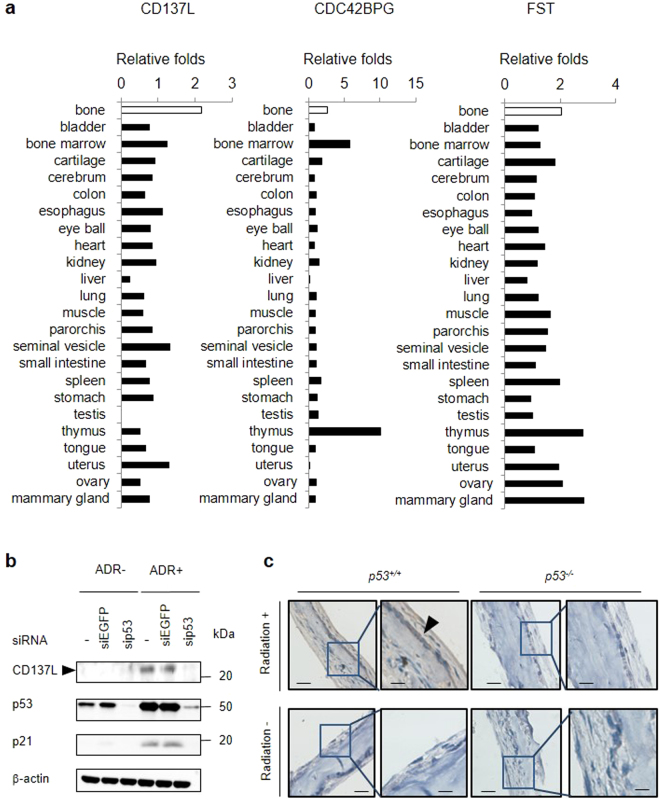
Figure 3.
Identification of CD137L as a bone-specific p53 target gene. (a) Induction of Cd137l among 24 tissues. Samples were categorized into 4 groups: (K) p53 −/− mice without irradiation, (W) p53 +/+ mice without irradiation, (KX) p53 −/− mice with irradiation and (WX) p53 +/+ mice with irradiation (n = 3 per group). We calculated the median FPKM value of WX/maximum FPKM value of the median FPKM value in K, KX or W for 24 tissues. (b) At 24 h after transfection with each siRNA, U2OS cells were treated with ADR (2 μg/ml for 2 h). At 36 h after treatment, whole cell extracts were subjected to western blotting with an anti-CD137L, anti-p21, anti-p53, or anti-β-actin antibody. siRNA against EGFP was used as a control. β-actin was used for normalization of the expression levels. These images were cropped from full-length blots (Supplementary Fig. 9). (c) Immunohistochemical staining of Cd137l in mouse p53α or p53−/− calvaria with or without radiation exposure. Representative images from three tissues in each group are shown. Scale Bars (left) = 50 μm, Scale Bars (right) = 20 μm. Black arrowhead shows osteoblast.

