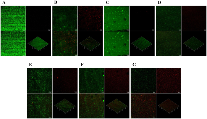
Figure 6.
Viability of biofilm in presence of 16~256 µg/mL of methyl gallate. The confocal microscopic images of LIVE/DEAD stained biofilm in (A) drug free control; biofilm treated with (B) furanone 10 µg/mL; (C) 16 µg/mL, (D) 32 µg/mL, (E) 64 µg/mL, (F) 128 µg/mL, and (G) 256 µg/mL of methyl gallate. The viability of the biofilms was assessed using BacLight LIVE/DEAD stain (green: live cells, red: dead cells). In each image, the segment at below right side shows three dimensional and other three segments shows two dimensional images.

