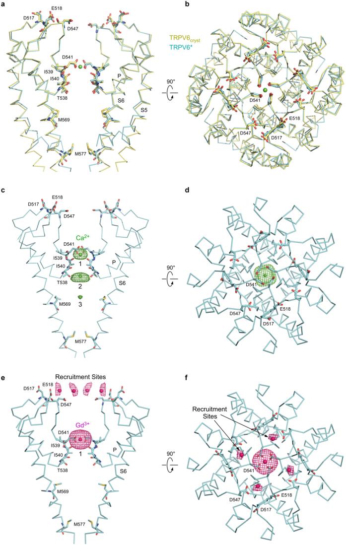
Figure 6.
TRPV6 structures with swapped and non-swapped transmembrane domains have identical ion channel pores. (a,b) Superposition of the pore domains in TRPV6* (cyan) and TRPV6cryst (yellow) viewed parallel to the membrane (a) or extracellularly (b), with residues important for cation binding shown in stick representation. Calcium atom is shown as a green sphere. (c–f) Side (c,e) and top (d,f) views of the TRPV6* pore. Front and back subunits in c and e are removed for clarity. Green and pink mesh shows anomalous difference electron density for Ca2+ (c,d, 3σ) and Gd3+ (e,f, 3.5σ), respectively, and ions are shown as spheres of the corresponding color. Positions of cation binding sites are similar to TRPV6cryst 5.

