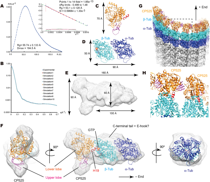
Figure 5.
Solution structure of CRMP2-GTP-tubulin hetero-trimer complex from small angle X-ray scattering. (A) Pair distance distribution functions (P(r)) and the Guinier plot. (B) Agreement of scattering data with GASBOR models (χavg = 2.21 ± 0.082). (C,D) Ribbon models of CRMP2 solved (C) and tubulin-dimer (PDB ID = 1JFF) (D) with size indication. (E) SAXS envelope for the CRMP2-tubulin complex. (F) Fitting of atomic models of CRMP2 and tubulin-dimer into the SAXS envelope. (G) Fitting of SAXS model of CRMP2-tubulin complex into the plus-end of the microtubule with 13 protofilaments (EMD-5195). (H) Close-up view of the dotted square in panel (G) depicts no steric clashes between neighboring CRMP2s. Data are mean ± s.d.

