Figure 7.
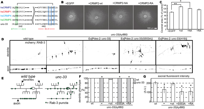
Two interface mutants affect CRMP2 functions in chick and c-elegans neurons. (A) Sequence alignment of the regions to be mutated. T246-N247, green; K483–R485–R487, blue. N247 hydrogen-bonds with S486 in the wild type CP525 structure. (B) Micrograph of DRG explants. Chick E7 DRG explants were infected with recombinant herpes-simplex virus harboring the indicted constructs (see Fig. S4 for infection efficiency). The neurites were visualized by α-tubulin immunostaining. Note that CRMP2-wild type (wt) enhances the neurite-outgrowth from the explant. This effect is canceled by the expression of RA mutant. Scale bar, 500 µm. (C) Scored graph. The covered area with the neurites was measured with ImageJ software. The relative area size of each sample was scored against the average of EGFP controls in the same experiment. Bar graph represents the mean ± s.e.m of 24 samples in each condition from 4 independent experiments. *P < 0.05; **P < 0.01, one-way ANOVA with post-hoc Tukey-Kramer test. (D–G) UNC-33 and UNC-33 mutants were expressed in unc-33(ky880): kyIs445 and the localization of mCherry::RAB-3 was observed in PVD neurons. (D) The localization of synaptic vesicles visualized by mCherry::RAB-3 in PVD neurons. wt, unc-33(ky880) and unc-33(ky880) expressing UNC-33 mutants were observed in the dendritic region and the axonal region. (E) Schema showing the localization of synaptic vesicles in wt and unc-33. (F) Dendritic mislocalization was statistically analyzed. The phenotype was sorted to either wt or unc-33 and counted. *P < 0.01, compared to wt, Chi-square test, n = 100 worms. (G) The axonal fluorescent intensity was measured in each strain. Mean ± S.D., *P < 0.01, compared to control, Dunnett test, n = 20 worms.

