Figure 1.
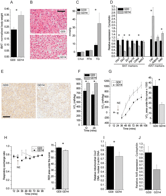
Pregnancy mediates changes in the morphometry and function of BAT. (A) Interscapular BAT (iBAT) weight expressed as a percent of total body weight without uterine horn at gestational days 0 (GD0) and 14 (GD14). (B) H&E staining of iBAT. Scale bar, 100 µm. (C) Concentrations of total cholesterol (Chol), free fatty acids (FFA) and triglycerides (TG), normalised to protein, in iBAT. (D) Expression of genes related to BAT and WAT phenotype. (E) Immunohistochemical staining for UCP1 in iBAT. Scale bar, 100 µm. (F) Basal measurement of O2 consumption in terminally anaesthetised mice. (G) Measurement of norepinephrine (NE)-mediated O2 consumption in terminally anaesthetised mice over time with O2 consumption total area under the curve. To facilitate comparisons, responses are expressed as increases over pre-NE values. (H) NE-mediated changes in respiratory exchange ratio (RER) over time with RER total area under the curve. (I) qPCR analysis of DNA levels of mitochondrial Cox2 normalised to nuclear β-actin in iBAT. (J) Expression of the mitochondrial-encoded gene Nd5 in iBAT.

