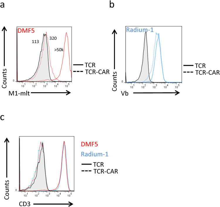
Figure 2.
Membrane expression of TCR-CAR. (a) DMF5 TCR and TCR-CAR were expressed in J76 cell line. Forty-eight hours later, cells were stained with pMHC multimers of HLA-A2 in complex with MART-1 peptide (M1). Mock transduced cells (grey) were used as negative control, TCR and TCR-CAR (red, plain and dotted, respectively) are shown. Numbers specify MFI of the indicated staining. (b) Same as in (a) but Radium-1 TCR (plain) and TCR-CAR (dotted) were here expressed and J76 cells were stained with anti-Vb3 antibody (Vb). (c) The cells as in A (red) and B (blue) were stained with anti-CD3 antibody. As before, TCRs are shown as plain lines and TCR-CAR as dotted, and mock transduced is in grey. These are representative staining of similar experiments performed on different retroviral preparation of cells at least once.

