
Fig. 1.
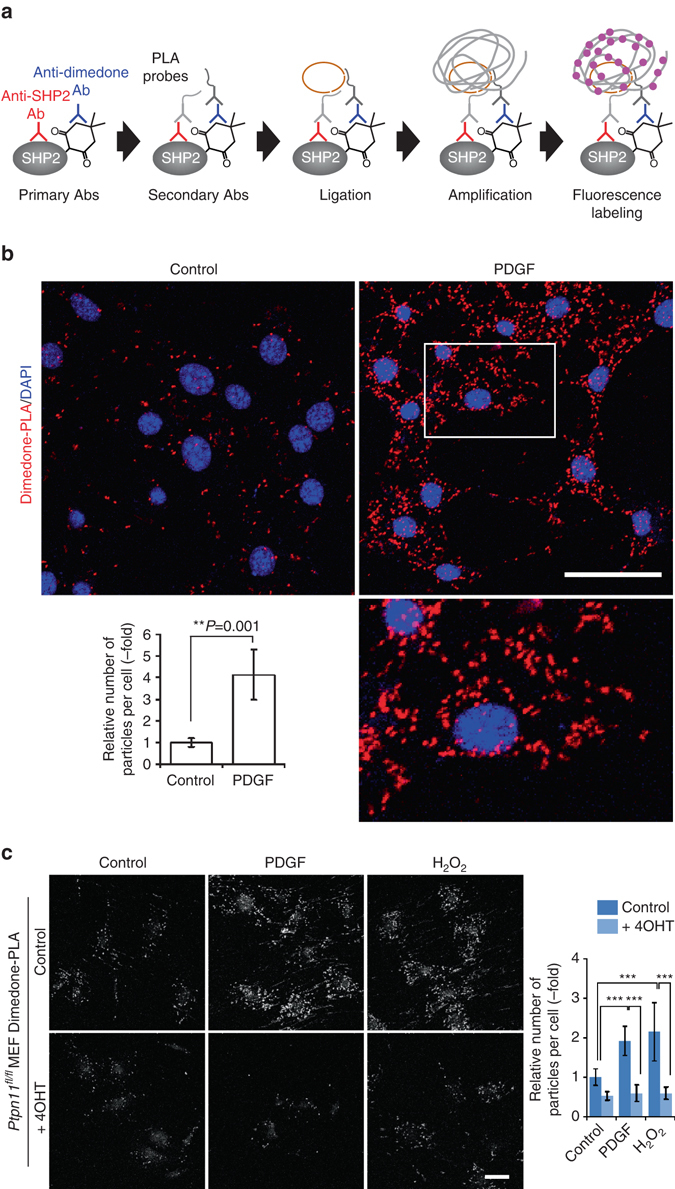
Detection of oxidized SHP2 by dimedone-PLA. a Schematic illustrating method for detecting oxidized SHP2 by dimedone-PLA. b Serum-starved Swiss 3T3 cells were stimulated with PDGF-BB (50 ng ml−1) for 10 min or left unstimulated. Cells were fixed in the presence of dimedone (5 mM) for 5 min, and subjected to dimedone-PLA (red). Nuclei were stained with DAPI (blue). Representative images are shown for each condition from one of >4 independent biological replicates. A higher magnification image of the boxed region is shown at the bottom right. The graph shows the average number of PLA signals per cell (n = 6 images for each condition, 5–20 cells in an image), relative to control cells without stimulation (set as 1). The P-value was calculated using a two-tailed Welch’s t-test. Error bars represent SD. c Serum-starved Ptpn11 fl/fl MEFs expressing CRE-ERTam treated with or without 4-hydroxytamoxifen (4OHT) were stimulated with PDGF-BB (50 ng ml−1) or H2O2 (1 mM) for 10 min. Cells were fixed in the presence of dimedone, and subjected to dimedone-PLA (gray). Representative images are shown for each condition from one of three independent experiments. The graph shows average number of PLA signals per cell (n = 6 images for each condition, 5–20 cells in an image), relative to control cells without stimulation (set as 1). ***P < 0.0001, ANOVA with Bonferroni/Dunn’s post-hoc test. Error bars represent SD. Scale bars: 50 μm