
Fig. 2.
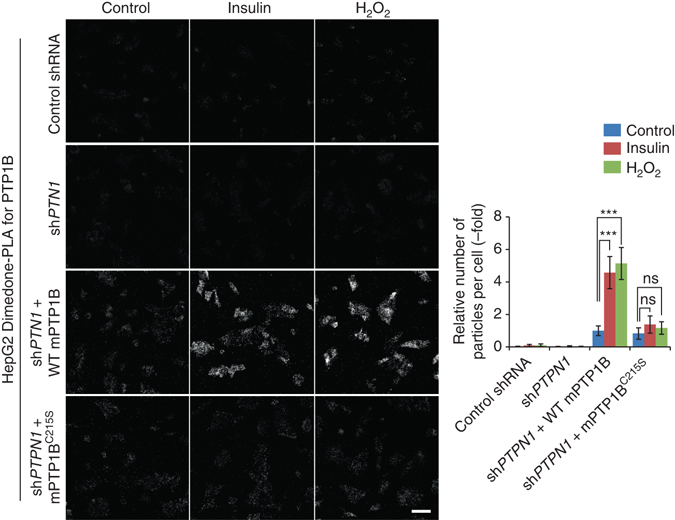
Detection of oxidized PTP1B by dimedone-PLA. Serum-starved HepG2 cells expressing control shRNA or shRNA targeting human PTPN1 (shPTPN1), with or without stable expression of either mouse WT PTP1B or PTP1BC215S, were stimulated with insulin (25 nM) or H2O2 (1 mM) for 5 min or left unstimulated. Cells were then subjected to dimedone-PLA with anti-dimedone-Cys and anti-mouse PTP1B antibodies (gray). Representative images are shown for each condition from one of two independent biological replicates. The graph shows the average number of PLA signals per cell (n = 6 images for each condition, 80–120 cells in an image), relative to control cells without stimulation (set as 1). ***P < 0.0001, ns not significant, ANOVA with Bonferroni/Dunn’s post-hoc test. Error bars represent SD. Scale bar: 50 μm