Figure 1.
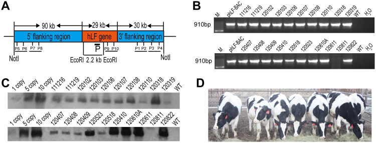
Generation of marker-free hLF BAC transgenic cloned cows. (A) Schematic representation of the transgene. The hLF BAC containing the complete hLF genomic DNA (a 29 kb genomic sequence consisting of human hLF flanked by a 90 kb 5′ flanking region and a 31 kb 3′ flanking region) was nucleofected into BFFs without a marker gene. The black box shows the 600 bp probe in Southern blotting upon digestion with EcoRI and a band of 2.2 kb resulting from the transgenic cloned cows. P1–P10 are five pairs of primers in different BAC regions for the identification of the transgenic cloned cows. (B) PCR analysis of transgenic cloned cows. Primers P1 and P2 amplified a 910 bp product to confirm positive transgenic cloned cows. M, 100 bp DNA ladder; pBAC-hLF, positive control; lanes 3–12, genomic DNA from transgenic cloned cows; H2O and WT, negative controls. (C) Southern blot identification of transgenic cloned cows. When digested with EcoRI, a band of 2.2 kb is detected in transgenic cloned cows. Lanes 1–3, positive controls with 1, 5 and 10 copies; Lanes 4–12, genomic DNA from transgenic cloned cows; WT, wild-type cow. (D) Image of transgenic cloned cows. The image shows transgenic cloned cows at the age of 10 months.

