Figure 2.
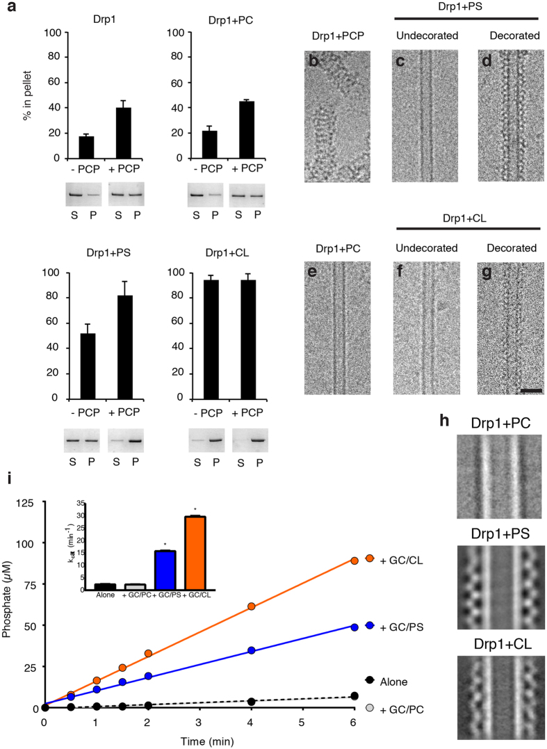
Drp1 recruitment and activation is enhanced with cardiolipin (CL) nanotubes. (a) Sedimentation analysis are presented for Drp1 alone, incubated with phosphatidylcholine nanotubes (GC/PC), phosphatidylserine nanotubes (GC/PS) and cardiolipin nanotubes (GC/CL) in the absence and presence of GMPPCP (−PCP and +PCP, respectively, n = 3/sample. Representative supernatant (S) and pellet (P) fractions are shown. Cryo-EM images are shown of Drp1 in the presence of GMPPCP (b), PS nanotubes (undecorated, (c), and decorated, (d)), PC nanotubes (no protein decoration observed, (e)), and CL nanotubes (undecorated, (f), and decorated, (g)). Scale bar, 50 nm. (h) 2-D class averages of Drp1 + GC/PC, Drp1 + GC/PS and Drp1 + GC/CL are presented. (i) A GTP hydrolysis assay displays the amount of phosphate released over time for Drp1 alone (black) and incubated with different nanotubes (GC/PC, gray; GC/PS, blue; GC/CL, orange, n = 3/sample). Measured GTPase activities (kcat) are shown (inset).

