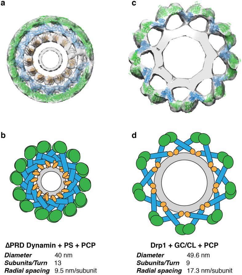
Figure 5.
Comparison of the lipid-bound Dynamin and Drp1 polymers. (a) The 3D cryo-EM reconstruction of the ΔPRD dynamin helical oligomer formed in the presence of PS liposomes and GMPPCP is presented with the GTPase (green), stalk (blue) and PH (yellow) domains fitted16. (b) The tight helical packing of dynamin is illustrated and the helical parameters are shown. (c) The 3D cryo-EM reconstruction of the Drp1 helical oligomer formed in the presence of CL-containing nanotubes and GMPPCP is presented with the GTPase (green) and stalk (blue) domains (PDB ID: 4BEJ) fitted. (d) The more expanded helical packing of the Drp1 helix is illustrated and the helical parameters are shown.

