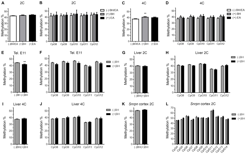
Figure 6.
Reduced methylation levels in the PCD of 4C cortical neurons seems to be partially due to the presence of 5fC. (A) Global pyrosequencing analysis of the CpG sites present in the PCD, performed with untreated (white), BH- (gray), or EA-treated genomic DNA (black) from 2C cortical neurons. (B) Pyrosequencing analysis of individual CpG sites (numbered as in Figure 2A), performed with untreated (white), BH- (gray), or EA-treated genomic DNA (black) from 2C cortical neurons. (C) Global pyrosequencing analysis of the CpG sites present in the PCD, performed with untreated (white), BH- (gray), or EA-treated genomic DNA (black) from 4C cortical neurons. (D) Pyrosequencing analysis of individual CpG sites (numbered as in Figure 2A), performed with untreated (white), BH- (gray), or EA-treated genomic DNA (black) from 4C cortical neurons. (E, G and I) Global pyrosequencing analysis, performed with genomic DNA treated either with BH (black) or left untreated (gray), of the CpG sites present in the PCD of E11 telencephalic neuroepithelium (E), 2C hepatocytes (G), and 4C hepatocytes (I). (F, H and J) Pyrosequencing analysis, performed with genomic DNA treated either with BH (black) or left untreated (grey), of individual CpG sites (numbered as in Figure 2A) present in the PCD of E11 telencephalic neuroepithelium (F), 2C hepatocytes (H) and 4C hepatocytes (J). (K) Global pyrosequencing analysis, performed with genomic DNA treated either with BH (black) or left untreated (grey), of the CpG sites present in the SCD of 2C cortical neurons. (L) Pyrosequencing analysis, performed at P15 with genomic DNA treated either with BH (black) or left untreated (grey), of individual CpG sites (numbered as in Figure 2B) present in the SCD of 2C cortical neurons. **P < 0.01.

