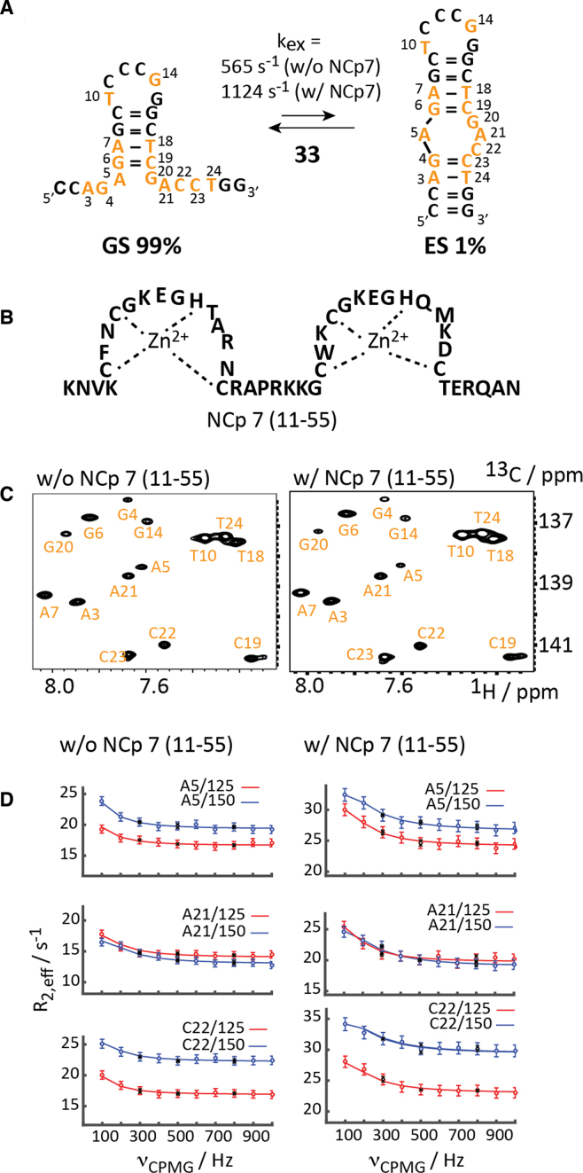
Figure 3.
13C-CPMG relaxation dispersion NMR of mini-cTAR DNA. (A) Exchange process for the site-specific 13C-modified mini-cTAR DNA. 13C-modified residues are highlighted in orange. (B) Schematic representation of the zinc-finger NCp7 peptide (11–55). (C) 1H-13C-HSQC spectra in the absence and presence of one equivalent NCp7 (11–55). (D) Non-flat carbon relaxation dispersion profiles of residues A5, A21 and C22 with and without NCp7 (11–55). Dots represent experimental data, lines the best fit, black crosses repeat experiments. Residue number and field strengths are specified.

